Hawkes, P.W. (Hrsg.)
Spence, John C.H. (Hrsg.)
Science of Microscopy

Beschreibung
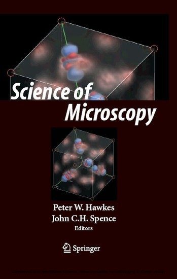
The examination of structure at the microscopic scale, between micrometers and angstrom units, has changed dramatically in recent decades. Many new types of microscopy have emerged, notably the many scanning-probe designs, some of which also allow manipulation of atoms to form wanted structures, while others now permit direct observation of moving proteins in liquids. The traditional electron microscope is being revolutionized by the arrival of aberration correctors and monochromators, which bring the resolution below the Angstrom and electron-volt level. The "laboratory in a microscope" concept is rapidly evolving, as nanostructures are observed forming under controlled conditions at atomic resolution (the carbon nanotube being the most famous recent example). Electron holography and scanning transmission electron microscopy have become indespensable tools of the semiconductor industry. The oldest form of microscopy, optical microscopy, has been rejuvinated by the development of flourescent, confocal, and two-photon varients. Analytical Scanning X-ray microscopes and Photoemission microscopes at synchrotons now routinely provide spatially resolved electronic structure maps. Tomographic imaging has vastly increased the information content of practically all forms of microscopy, as reflected in the award of a recent Nobel prize. Molecular biology is benefitting enormously from progress in this technique. Most of these developments are responses to the urgent needs of researchers to characterize new useful nanostructures at the atomic level.
Produktdetails
| ISBN/GTIN | 978-0-387-49762-4 |
|---|---|
| Seitenzahl | 748 S. |
| Kopierschutz | mit Wasserzeichen |
| Dateigröße | 56750 Kbytes |

