Csillag, András (Hrsg.)
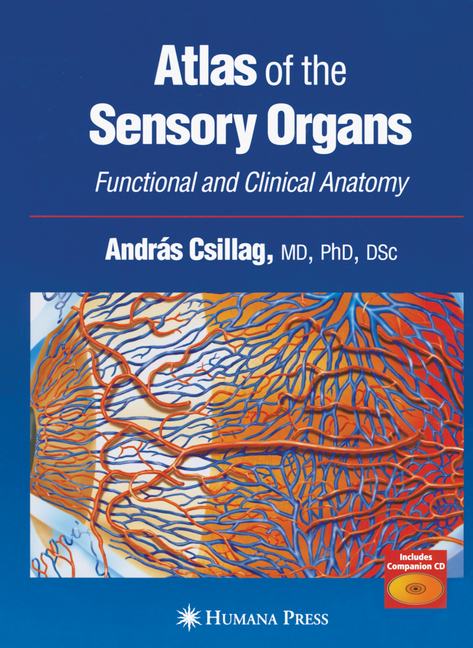
Atlas of the Sensory Organs
Functional and Clinical Anatomy

Beschreibung
A richly illustrated medical atlas of the five main human sensory systems together with their neural pathways, from primary sensation to processing by the brain. The authors provide a detailed anatomical survey of each sensory organ, covering their ontogeny (development), central pathways, and functional mechanisms. Highlights include microanatomy and endoscopic images of the temporal bone, human embryonic specimens demonstrating the histology of the developing ear, and scanning electron micrographs of the organ of Corti and the vestibular receptors. There are also easy-to-use tables providing an overview of the nerves and arteries of the eye and orbit and clinical specimens of the eye and optic pathways. A companion compact disc contains high resolution copies of the color illustrations used in the book. Although the sensory organs-particularly the eye and ear-are of great clinical importance, the lecture time allocated to them in the anatomical curricula is limited because of their great nomenclatural and physiological complexity. Sufficiently detailed descriptions can usually be found only in multivolume handbooks or in specialist monographs covering a single sensory organ. In Atlas of the Sensory Organs: Functional and Clinical Anatomy, a panel of expert medical educators and internationally renowned comparative and developmental neuroanatomists concisely, but thoroughly, describe the five main human sensory systems together with their neural pathways, from primary sensation to processing by the brain. The authors provide a detailed anatomical survey of each sensory organ, covering their ontogeny (development), central pathways, and functional mechanisms. The organs themselves are richly illustrated with light and electron microscopic representations of healthy and intact organs or tissues, except where pathology is directly relevant to understanding normal structure and function. Highlights include microanatomy and endoscopic images of the temporal bone, human embryonic specimens demonstrating the histology of the developing ear, and scanning electron micrographs of the organ of Corti and the vestibular receptors. There are also easy-to-use tables providing an overview of the nerves and arteries of the eye and orbit, and clinical specimens of the eye and optic pathways. A companion compact disk contains high-resolution copies of the color illustrations used in the book.
Produktdetails
| ISBN/GTIN | 978-1-59259-849-6 |
|---|---|
| Seitenzahl | 258 S. |
| Kopierschutz | mit Wasserzeichen |
| Dateigröße | 61388 Kbytes |

